Diagnostics · CBCT / DVT
Seeing the jaw in three dimensions.
Low-dose 3D imaging gives us a true volumetric view of bone, nerves and hidden inflammation - so treatment is planned on certainty, not on a flat shadow.
01 What it is
From a flat shadow to a real volume.
A conventional dental X-ray is a single, flattened image: structures sit on top of one another, and a great deal stays hidden behind them. Cone beam computed tomography - CBCT, in German DVT - works differently. The scanner makes one quiet rotation around your head and captures a three-dimensional volume of your jaw in seconds.
That volume can then be examined slice by slice and from any angle. We can measure the exact height and width of bone, follow the true course of a nerve, assess the maxillary sinus, and look into areas of the jawbone that a flat picture simply cannot reveal. The diagnosis stops being an educated guess and becomes a measurement.
- A genuine 3D view of jaw, teeth, nerve canals and sinuses.
- Captured in a single, low-dose scan lasting only seconds.
- Reviewed from any angle, with precise distances and bone density.
- The foundation for safer, gentler, better-planned treatment.
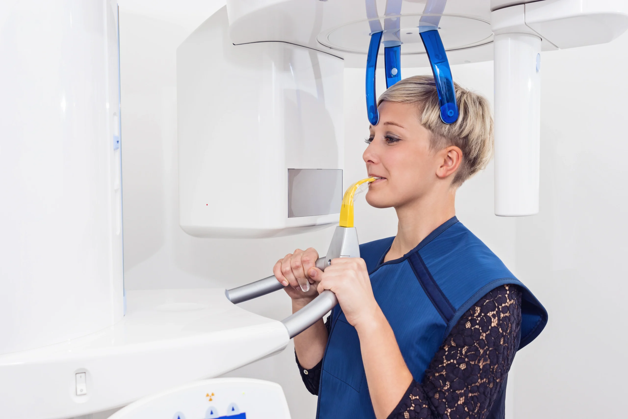
Fig. 01A reconstructed CBCT volume of the jaw, viewed in our practice near Lübeck.
02 Low dose, used deliberately
The most information,
for the least exposure.
A dental CBCT delivers far less radiation than a medical CT of the same region - and we take one only when a flat image cannot answer the question. The field of view is kept as small as the diagnosis allows. That is the ALARA principle: as low as reasonably achievable.
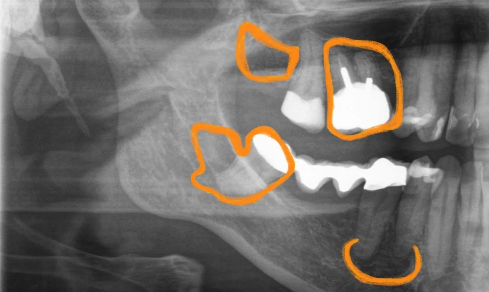
Fig. 02Evaluating bone density where silent inflammation can hide.
03 Finding what stays hidden
Silent inflammation - NICO and FDOK.
Some of the most important findings in biological dentistry are the ones that never hurt. FDOK (fatty-degenerative osteonecrosis of the jaw) and NICO describe chronic, poorly healed areas of bone - often at the site of a long-ago extraction. They are usually painless, and on a flat X-ray they hide easily behind overlapping structures.
In a whole-body view of health, these quiet areas matter: they can act as ongoing interference fields. A 3D scan lets us evaluate bone density in these regions in detail - which is exactly why three-dimensional imaging sits at the heart of a careful biological assessment rather than at its edge.
04 Precise implant planning
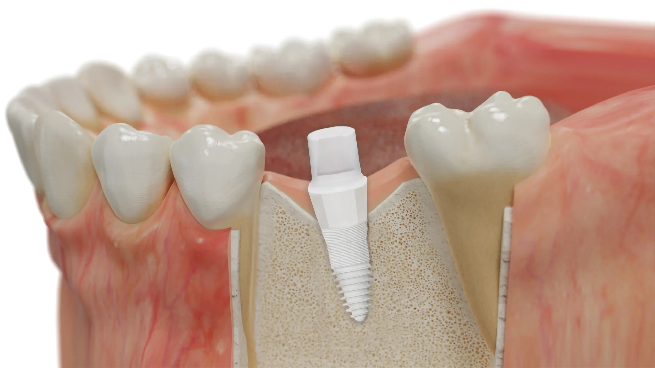
Planning a ceramic implant before we begin.
Placing a metal-free ceramic implant is, above all, a question of millimetres. How much bone is really there? Where exactly does the nerve run? How far is the sinus? A 3D dataset answers each of these in advance, in three dimensions, so the position and size of the implant are decided before any treatment starts.
The result is surgery that is more predictable and gentler - a safe distance kept from sensitive structures, and a plan made around your individual anatomy rather than an average one.
- Bone height and width measured precisely in three dimensions.
- A safe, planned distance from nerves and the maxillary sinus.
- Implant position and size chosen before any incision.
- More predictable, less invasive ceramic implant treatment.
05 In the practice
See it explained in English.
A short film on how we use low-dose 3D diagnostics to find hidden problems and plan treatment with precision.
06 What to expect
A 3D scan, from start to finding.
A real indication
We take a 3D scan only when a flat image cannot answer the clinical question - never routinely.
The scan itself
You sit or stand still while the scanner makes one quiet rotation. The capture takes only seconds.
Careful review
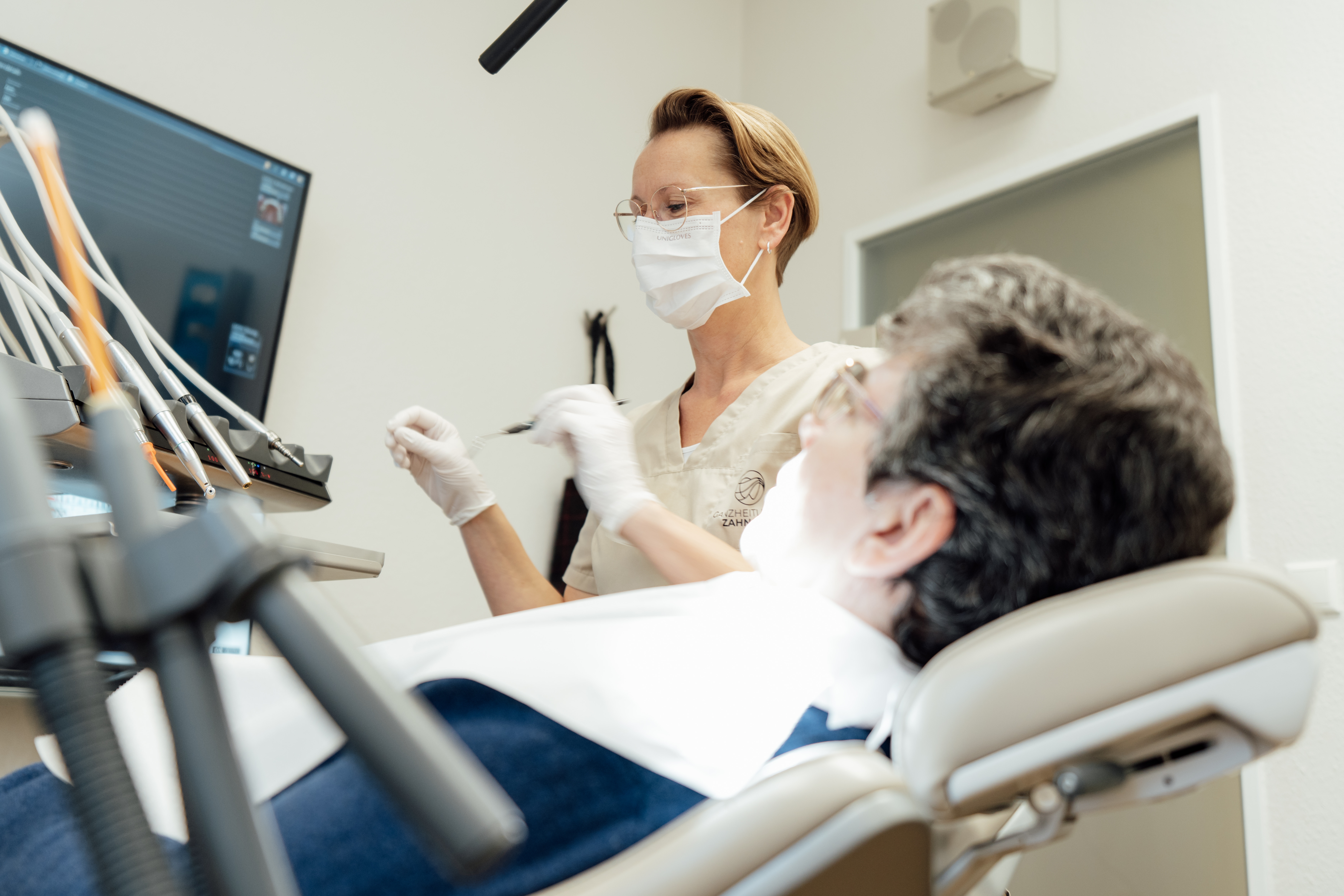
The volume is examined slice by slice - bone, nerves, sinus and any silent inflammation.
Your findings, explained
We talk you through what the scan shows - in English - and what it means for your treatment plan.
07 When 3D imaging helps
Where a three-dimensional view changes the answer.
3D imaging is not for every visit - but in these situations it turns guesswork into a clear picture. What the scan involves financially is covered in our guide to CBCT scan cost.
Before a ceramic implant
Measuring bone in three dimensions and keeping a safe distance from nerves and the sinus.
02Searching for hidden inflammation
Evaluating possible NICO / FDOK and chronic interference fields as part of a whole-body assessment.
03Complex or unclear findings
Difficult root anatomy, impacted teeth or the assessment of older treatments that a flat image leaves ambiguous.
08 Questions
3D imaging, answered plainly.
A dental cone beam CT delivers far less radiation than a medical CT of the same region, because only the jaw is imaged and the field of view is kept as small as the question allows. We follow the ALARA principle - as low as reasonably achievable - and take a 3D scan only when a two-dimensional image cannot answer the clinical question.
A conventional X-ray flattens everything onto one plane, so structures overlap. A CBCT volume can be viewed from any angle, so we can measure bone height and width, see the exact path of nerves, assess the sinus, and detect hidden inflammation in the jawbone that a flat image easily misses.
FDOK (fatty-degenerative osteonecrosis of the jaw) and NICO describe chronic, often silent areas of poorly healed bone in the jaw - frequently at former extraction sites. They rarely hurt and are hard to see on a flat X-ray. A 3D scan lets us evaluate bone density in these areas in detail, which is why it is central to a biological, whole-body assessment.
For implant planning a 3D scan is extremely valuable. It lets us measure the available bone in three dimensions, keep a safe distance from nerves, and choose the position and size of a metal-free ceramic implant before any treatment begins - which makes surgery more predictable and gentler.
Yes. The practice in Bad Schwartau near Lübeck welcomes English-speaking and international patients, and we explain your 3D findings and the resulting plan in English.
Plan your treatment on certainty.
Tell us about your situation. We reply personally - in English - and let you know whether a 3D scan would help.
Request an appointment