Biological surgery · FDOK / NICO
FDOK / NICO: carefully assessing a suspected, often silent jawbone finding.
FDOK - known in English as NICO - describes a suspected, debated finding: chronic, often painless changes in the jawbone. Because such areas are hard to judge on flat X-rays, we assess them in three dimensions - and only where a well-founded finding is confirmed do we treat it gently, biologically.
Phone hours: Mon–Thu 8:30–13:00 (German time)
01 What it is
What FDOK / NICO describes.
FDOK and NICO describe the same suspected finding: chronic, aseptic changes in the bone marrow of the jaw that, in the biological-dentistry model, are discussed as a quiet source of inflammatory signalling molecules. According to this model, the bone does not fully heal after an earlier injury or extraction, and a soft, fatty, poorly mineralised area may be left behind - often without any pain to announce it.
In a whole-body view of health, these quiet zones are taken seriously. In the biological-dentistry model they are described as possible ongoing interference fields that some practitioners consider may release inflammatory messengers such as RANTES/CCL5 into the body - a viewpoint rather than an established medical fact. That is why, in biological dentistry, we look at them carefully even when nothing hurts.
FDOK
Fettig-degenerative Osteonekrose des Kieferknochens - fatty-degenerative osteonecrosis of the jawbone.
NICO
Neuralgia-Inducing Cavitational Osteonecrosis - the name more often used in English literature.
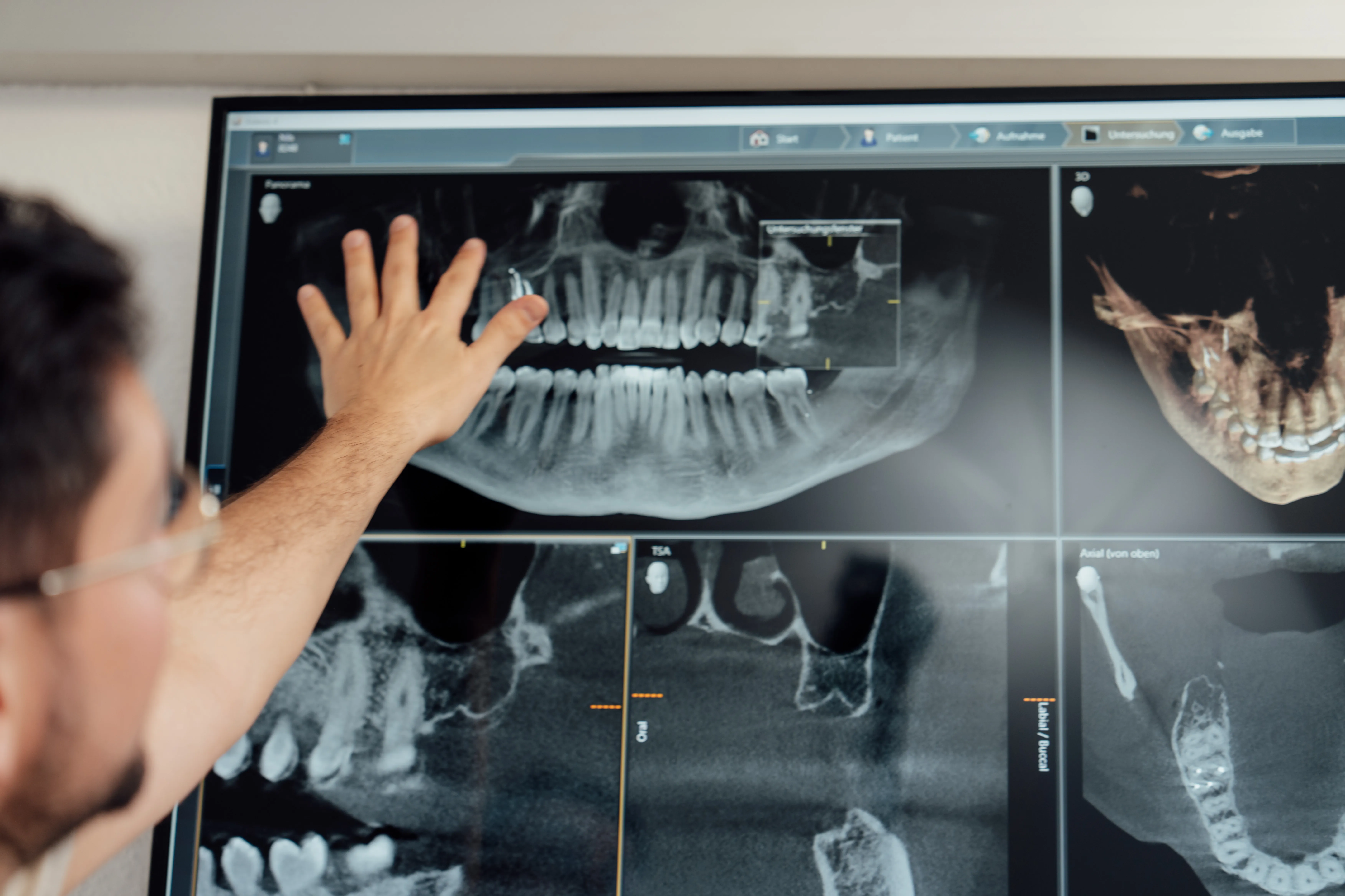
Fig. 01Reviewing jaw imaging - where silent areas first become visible.
02 Names & evidence
FDOK, NICO, cavitation – three names, one debate.
The same idea travels under several names. In German-speaking biological dentistry it is called FDOK – fettig-degenerative Osteolyse/Osteonekrose des Kieferknochens, a fatty-degenerative breakdown of the jawbone. In English-language literature the term NICO – neuralgia-inducing cavitational osteonecrosis – goes back to descriptions of hollow, poorly healed bone associated with facial pain. In the United States, patients mostly meet the everyday term jawbone cavitation. All three point to the same suspected finding: an area, often at an old extraction site, where the bone may not have healed into normal, well-mineralised tissue.
It is only fair to say that this condition is debated. The International Academy of Oral Medicine and Toxicology describes jawbone cavitations as a real disease in its IAOMT position paper on human jawbone cavitations and proposes the name “chronic ischemic medullary disease of the jawbone”. Many mainstream dental and pain specialists disagree: a 2025 review in the Journal of Pain Research concludes that jawbone cavities have not been validated as a distinct disease, that their role in facial pain remains unproven, and that properly designed clinical studies are needed.
What this means for you in practice: an assessment rests on your history and clinical findings together with 3D CBCT imaging. A CBCT can show areas of reduced bone density, but it has limits – on its own it does not prove FDOK / NICO, and findings have to be read with caution. Surgery is only considered when there is a well-founded finding, after other causes have been looked at, and after we have talked through the options, uncertainties and risks with you. Not every suspicion leads to treatment.
Unsure whether this applies to you? Describe your situation →
03 Why it matters
A quiet area of bone
can still be worth attention.
An FDOK / NICO site rarely causes local pain - yet in the biological-dentistry model it is discussed as something that may keep releasing inflammatory messengers and act as a lasting interference field. Looking at the mouth as part of the whole body is the heart of a biological approach: the aim is to address possible causes, not only symptoms. Outcomes vary from person to person and cannot be promised.
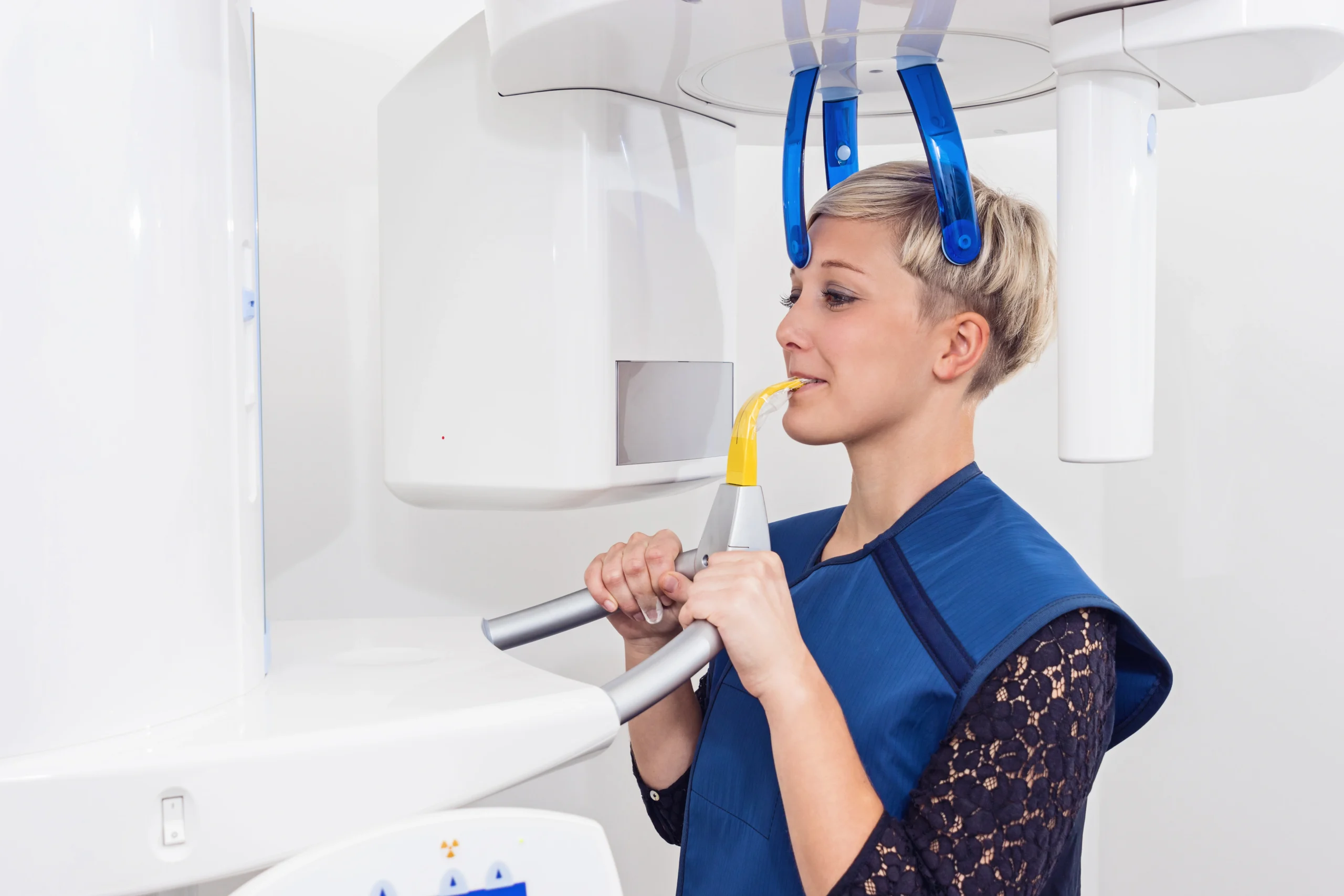
Fig. 02A 3D dataset reveals bone density that a flat image cannot.
04 How we find it
Diagnosis in three dimensions.
Suspected areas are notoriously hard to judge on standard two-dimensional X-rays - they can look unremarkable on a flat image because overlapping structures hide them. So we do not rely on a flat shadow.
Instead we combine 3D CBCT imaging, which lets us judge bone density from any angle, with a careful clinical assessment and, where indicated, blood analysis of inflammatory markers such as RANTES/CCL5 - which is not a validated diagnostic test for FDOK / NICO. Together these give a clearer picture before any treatment is considered - though even a 3D scan cannot prove FDOK / NICO on its own.
- Three-dimensional view of bone density, not a flat overlap.
- Clinical history correlated with the imaging findings.
- Blood markers (e.g. RANTES/CCL5) as supporting information only - not a validated test for FDOK / NICO.
05 The protocol
Gentle, biological surgery - step by step.
When treatment is the right choice, it follows a meticulous protocol, planned in advance from your 3D dataset.
Gentle removal
Where a well-founded finding is confirmed, the affected, poorly healed bone is carefully removed under local anaesthesia and the area thoroughly cleaned.
Ozone disinfection
The site is disinfected with ozone - a gentle approach intended to reduce bacteria without harsh chemicals.
PRF from your own blood
PRF membranes, prepared from a small sample of your own blood, are placed to support natural regeneration.
Calm recovery
Recovery varies; many people can return to normal activities within a few days. We explain aftercare in detail.
06 Why this way
Metal-free, body-first, fully explained.
Every choice in this protocol follows the same idea: work with the body rather than against it. We use no metal and no foreign growth additives - the regeneration is supported by your own blood in the form of PRF, and the disinfection is done with ozone rather than aggressive agents.
Just as importantly, you are never left guessing. We talk you through what the 3D scan shows, why treatment is or is not advised, and exactly what each step involves - in English, before anything begins.
- No metal, no foreign additives - a biological approach throughout.
- Regeneration supported by PRF from your own blood.
- Planned in advance from a precise 3D dataset.
- Findings and options explained clearly in English.
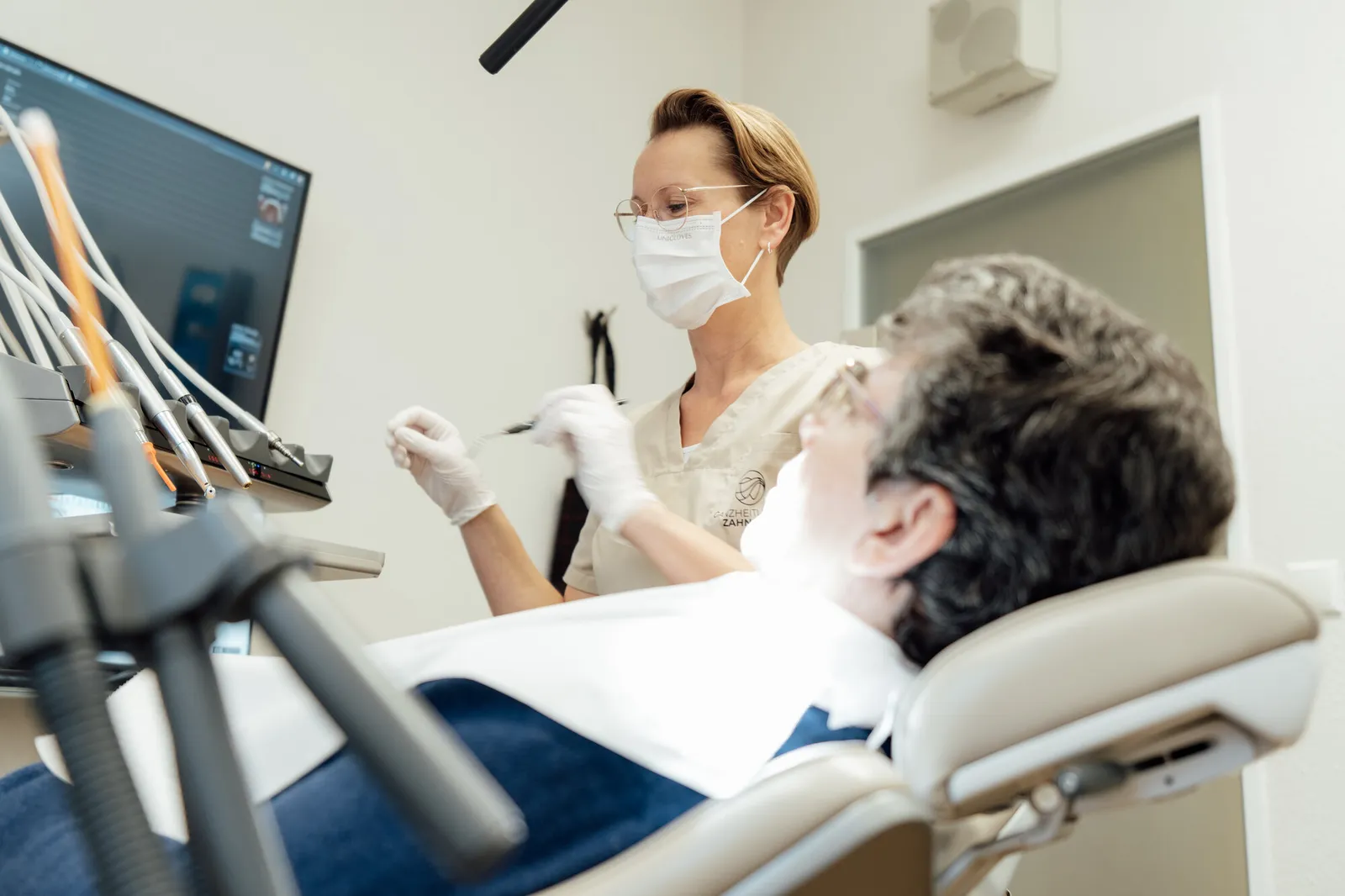
Fig. 03Treatment carried out gently, in our practice near Lübeck.
07 When to look closer
Who an FDOK / NICO assessment can help.
An assessment is worth considering when symptoms have no clear dental cause, or when a whole-body picture points back to the jaw.
Unexplained facial or jaw pain
Persistent neuralgia or discomfort that conventional dental findings cannot fully explain.
02Old extraction or root-canal sites
Areas where a tooth was removed long ago and the bone may not have healed completely.
03Search for possible interference fields
A possible chronic inflammatory load with no obvious source, considered as part of a whole-body biological assessment - a model used in biological dentistry rather than a confirmed diagnosis.
08 Questions
FDOK / NICO, answered plainly.
They are two names for the same suspected, debated finding. FDOK is the German term - fettig-degenerative Osteonekrose des Kieferknochens, fatty-degenerative osteonecrosis of the jaw. NICO is the term more common in English literature - Neuralgia-Inducing Cavitational Osteonecrosis. Both refer to suspected chronic, often silent areas of poorly healed bone in the jaw, typically at former extraction sites - a concept that mainstream dentistry has not validated as a distinct disease.
Broadly, yes - they are different names for the same suspected finding. FDOK is the German term (fettig-degenerative Osteolyse/Osteonekrose des Kieferknochens), NICO stands for neuralgia-inducing cavitational osteonecrosis, and jawbone cavitation is the everyday term used mainly in the US. The condition itself is debated: proponents such as the IAOMT regard it as a distinct disease, while many mainstream specialists consider it unproven. An assessment therefore combines your history, a clinical examination and 3D CBCT imaging - and the imaging alone is not proof.
Suspected areas are usually painless, and on a conventional two-dimensional X-ray they often look unremarkable because overlapping structures hide them. This is why we rely on three-dimensional CBCT imaging, combined with a careful clinical history and, where indicated, blood analysis of inflammatory markers such as RANTES/CCL5 - which is not a validated diagnostic test for FDOK / NICO. Even together, these findings cannot prove the condition on their own.
Treatment is only considered when there is a well-founded finding, and then follows a meticulous biological protocol. The affected bone is carefully removed under local anaesthesia, the site is disinfected with ozone, and PRF membranes prepared from the patient's own blood are placed to support natural regeneration. Each step is planned in advance from the 3D dataset.
PRF - platelet-rich fibrin - is prepared by spinning a small sample of your own blood. The resulting membrane is rich in your body's own growth factors and contains no foreign additives. Placed in the treated site, it is intended to support the natural healing of bone and soft tissue, which fits the metal-free, biological approach of the practice.
Recovery varies from person to person. The procedure is carried out as gently as possible, and many people can return to their normal activities within a few days. We explain aftercare clearly and remain available for any questions.
There is no flat price, because every jaw is different - the imaging, the number of affected sites and the scope of any biological surgery all vary from person to person. After your 3D scan and a personal examination you receive a written, itemised estimate for your own case, so you can decide with the full picture in front of you before anything begins. For the imaging part, see what drives the cost of a 3D CBCT scan. Reimbursement may be possible through a private (supplementary) dental insurance - please clarify this with your own insurer in advance. You receive a standard invoice, itemised by service. Whether a public health system contributes anything is decided solely by your own insurer or authority; we do not advise on this.
Yes. The practice in Bad Schwartau near Lübeck welcomes English-speaking and international patients. We explain the diagnosis, the imaging findings and every step of treatment in English.
A question about your own jaw that isn’t answered here? Ask us directly →
09 Related reading
The whole-body picture around the jaw.
Teeth and the immune system
How chronic inflammation at root-canal and extraction sites connects the mouth to the immune system - the oral-systemic link, explained responsibly.
iiTeeth and organs: the tooth-meridian chart
Which tooth is traditionally linked to which organ in biological dentistry - presented honestly as a model, not as anatomy.
iiiBiological wisdom tooth extraction
Careful, socket-preserving removal that helps extraction sites heal fully - the same sites where FDOK / NICO can later develop.
10 Get started
Wondering whether the jaw plays a role?
Describe your situation briefly in the form. We reply personally - in English or German - with the next steps; the examination, including 3D diagnostics, takes place at the practice.
Practice
Hauptstraße 50
23611 Bad Schwartau
Germany
Medical information notice. The descriptions of FDOK / NICO, interference fields and inflammatory markers on this page reflect concepts and viewpoints used within biological dentistry. They are offered for general information, are not universally accepted in conventional medicine, and are not a promise of diagnosis, healing or cure. Whether any assessment or treatment is appropriate can only be decided individually after a personal examination.