
01  Biological dentistry · Explainer

Teeth and organs: understanding the tooth-meridian chart.

You may have seen a chart that links each tooth to an organ - incisors to the kidneys, canines to the liver, wisdom teeth to the heart. It is one of the most searched ideas in holistic dentistry, and also one of the most misunderstood. Here is a calm, honest guide to where the chart comes from, the full mapping, and - just as importantly - what it does and does not mean.

FDI numberingThe international tooth system
EAV / Voll modelA traditional lens, not a law
Evidence-awareModels named as models
EN & DEInternational patients welcome

02  Where the idea comes from

An old map of the body, drawn through the mouth.

The tooth-organ chart has two roots. The first is the meridian theory of Traditional Chinese Medicine (TCM), which describes the body as criss-crossed by channels - meridians - said to carry energy between organs and the surface of the body. In that framework, each tooth sits on one of these channels and is grouped with the organs the channel is thought to serve.

The second root is more recent: in the mid-twentieth century the German physician Dr Reinhold Voll developed Electroacupuncture according to Voll (EAV), an electrodermal method that mapped specific teeth to specific organs and tissues. The familiar “odonton” chart that circulates online today largely descends from his work.

In practice the chart works by grouping rather than by single tooth. Most associations follow the type of tooth - incisors, canines, premolars, molars and wisdom teeth - because the original models placed whole groups on the same channel. That is why you rarely see a chart singling out one isolated tooth: the mapping is organised by region of the mouth and the meridian thought to pass through it. It is also why charts from different authors broadly agree on the big groupings while disagreeing on the details.

Both TCM and EAV are observational, traditional models - ways of organising experience that long predate modern imaging and laboratory science. They can be genuinely interesting, and many patients find them a helpful way to think about the body as a connected whole. They are not, however, proven anatomical wiring, and we present them here exactly as that: a lens, not a law.

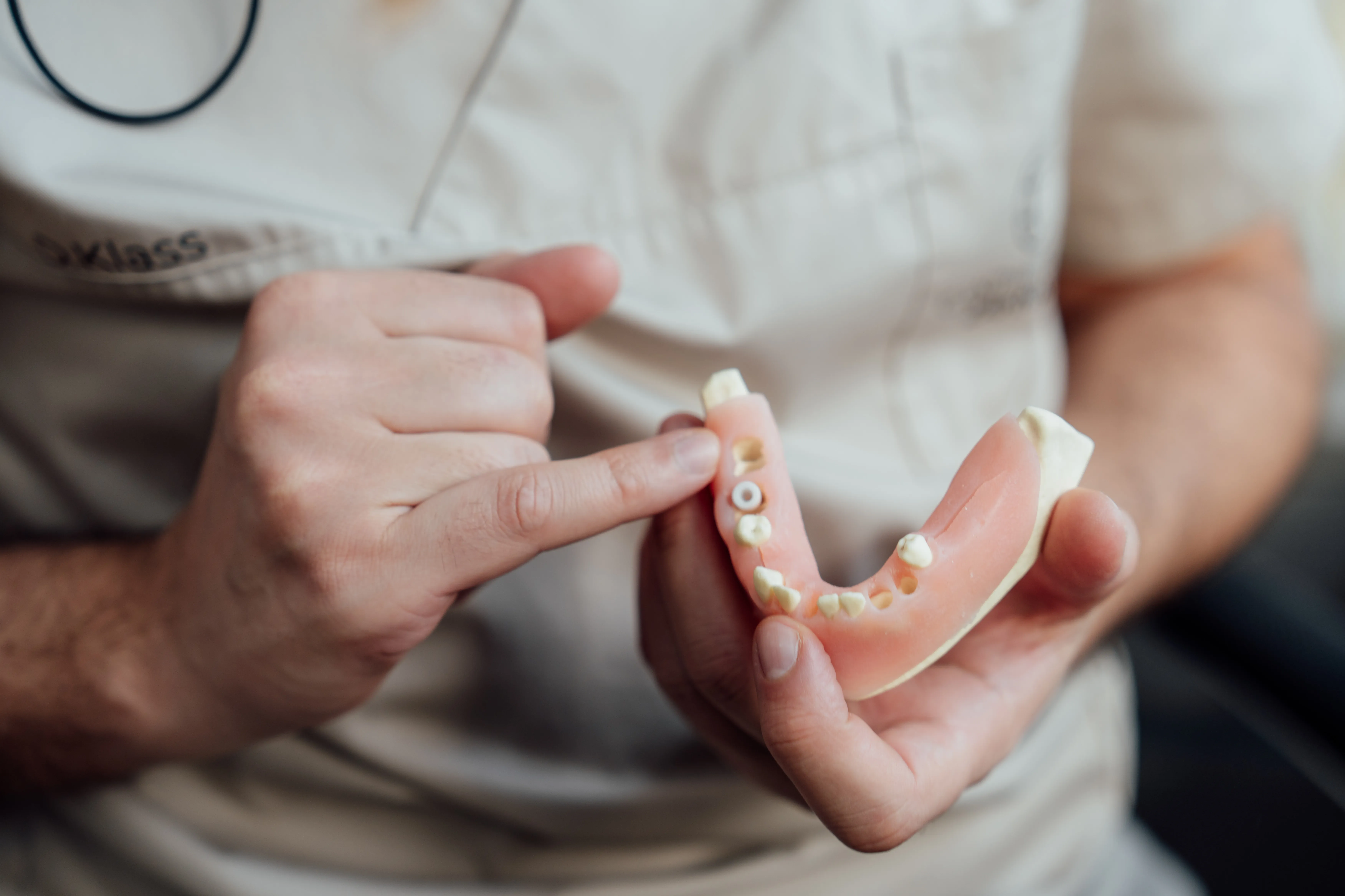
Calm biological dentistry consultation focused on whole-body health at the practice of Dmitri Klass in Bad Schwartau

Before we go further - a clear disclaimer. The tooth-meridian chart is an interpretive model that some biological dentists consider alongside conventional diagnostics. On this page it is not used to diagnose any illness, and it is not a promise that dental treatment will cure a bodily condition.

If a tooth and an organ share a meridian, that is a traditional association - not evidence that one is causing the other. Real decisions are always based on genuine clinical findings, and any health concern beyond the teeth belongs with your physician. We work with that boundary, not around it.

Natural, calm treatment environment at the holistic dental practice near Lübeck

03  Our position

Curious about the map.
Honest about its limits.

We find the tooth-organ chart a useful prompt for thinking about the whole person - and we refuse to dress a traditional model up as proven science. Both things can be true at once.

04  The chart in full

The tooth-organ-meridian table.

Here is the classic EAV/Voll mapping, written out with international FDI tooth numbers so you can find your own teeth. Read it from left to right: the tooth, its type, the meridian it is traditionally placed on, and the organs and tissues that meridian is associated with.

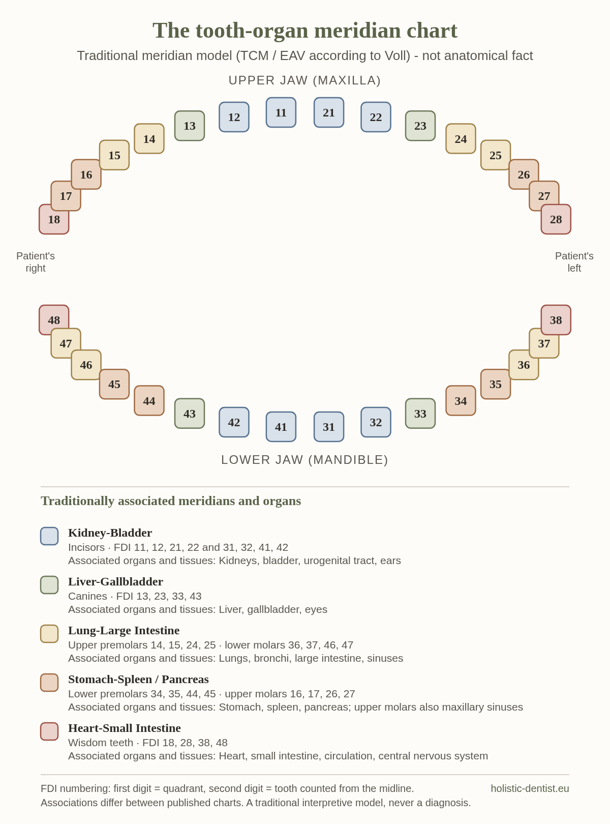
Tooth-organ meridian chart based on Traditional Chinese Medicine: which tooth is linked to which organ in meridian theory (FDI numbering)
The chart at a glance: both dental arches in FDI numbering, coloured by the meridian each tooth group is traditionally placed on. Traditional meridian model (TCM/EAV) - not anatomical fact. The table below spells out every row.
Traditional tooth-organ-meridian associations (EAV/Voll odonton model). FDI numbering. Published charts differ between sources - associations should be read as traditional, not anatomical.
Teeth (FDI) Tooth type Traditionally associated meridian Commonly associated organs & tissues
11, 12, 21, 2231, 32, 41, 42 Central & lateral incisors (upper & lower) Kidney-Bladder Kidneys, bladder, urogenital tract, ears
13, 2333, 43 Canines (upper & lower) Liver-Gallbladder Liver, gallbladder, eyes
14, 15, 24, 25 Premolars (upper) Lung-Large Intestine Lungs, large intestine, sinuses
34, 35, 44, 45 Premolars (lower) Stomach-Spleen / Pancreas Stomach, spleen, pancreas
16, 17, 26, 27 Molars (upper) Stomach-Spleen / Pancreas Maxillary sinuses, stomach, spleen, pancreas
36, 37, 46, 47 Molars (lower) Lung-Large Intestine Lungs, bronchi, large intestine
18, 28, 38, 48 Wisdom teeth / third molars Heart-Small Intestine Heart, small intestine, circulation, central nervous system

A practical note on reading the numbers: in the FDI system the first digit is the quadrant (1 = upper right, 2 = upper left, 3 = lower left, 4 = lower right) and the second is the tooth, counting from the midline. So “26” is the first upper-left molar. Associations differ between sources - you will find charts that place the upper molars or wisdom teeth slightly differently. That variation is itself a reason to treat the map gently.

A related idea: the tooth-vertebra chart

Alongside the organ chart, traditional models also link teeth to segments of the spine - a so-called tooth-vertebra chart. The reasoning is the same: each tooth group is mapped onto a region of the spinal column and its associated nerves. As with the organ map, this is a traditional correspondence rather than a demonstrated anatomical pathway. A back complaint is no more a dental diagnosis than a toothache is a spinal one.

We mention it because patients often find the two charts side by side online and assume that together they carry the weight of proof. They do not. They are the same kind of interpretive model, drawn from the same traditions, and they belong beside a proper examination - never in place of one.

05  Tooth by tooth

Which tooth is connected to which organ?

The table answers that in one view. Here it is written out the way people actually ask it - group by group, in plain sentences. Every association below is taken from the same traditional EAV/Voll mapping shown above; none of it is a diagnosis, and none of it says that a tooth makes an organ ill.

FDI 18 · 28 · 38 · 48 — wisdom teeth

Which tooth is connected to the heart?

The wisdom teeth - the third molars - are the teeth the chart places on the Heart-Small Intestine meridian, together with circulation and the central nervous system. That single row is the source of almost everything written online about teeth and the heart.

Why the wisdom teeth, of all teeth? Because of how the odonton model is built. Voll's chart does not wire single teeth to single organs; it places whole groups of the same tooth type on one channel, each tooth considered together with its root, the ligament that suspends it and the surrounding bone. The third molars form the last of those groups, and in his mapping that group landed on the Heart-Small Intestine line - the same row you can check in the table above. Other published charts keep the grouping but shift the details, which is one more reason to hold the row lightly.

Read it as a grouping in a traditional model. No nerve, vessel or duct runs from a wisdom tooth to your heart, and a symptom in the chest is never a dental question. If your heart concerns you, that belongs with your physician the same day - not after a dental appointment.

There is, separately, a documented side to teeth and the heart, and it has nothing to do with meridians. Chronic gum inflammation (periodontitis) is associated in the medical literature with cardiovascular disease - an association studied through inflammation, not proven causation. And for some heart patients, for example after endocarditis or a valve replacement, an antibiotic prophylaxis before invasive dental procedures is established medical practice, agreed with the treating physician. Those are the mouth-heart connections dentistry actually works with - described in the evidence-supported section below and in our approach to gum health.

FDI 11, 12, 21, 22 · 31, 32, 41, 42 — incisors

Which teeth are connected to the kidneys and bladder?

All eight incisors, upper and lower, sit on the Kidney-Bladder meridian. The same channel is traditionally associated with the urogenital tract and with the ears.

Because the incisors are the teeth people see every day, this is one of the most-searched rows of the chart - and one of the easiest to over-read. It tells you nothing about how your kidneys are working, and it is never a reason to treat a front tooth that is healthy.

FDI 13, 23, 33, 43 — canines

Which tooth is connected to the liver?

The four canines are grouped with the Liver-Gallbladder meridian, a channel the chart also associates with the eyes.

Liver function is something a blood test answers, not a canine. Where the association can be useful is as a prompt in conversation - if someone is already under medical care for their liver, we simply take extra care that nothing in the mouth is quietly adding an inflammatory load.

FDI 14, 15, 24, 25 · 36, 37, 46, 47 — upper premolars, lower molars

Which teeth are connected to the lungs?

Two groups share the Lung-Large Intestine meridian: the premolars of the upper jaw and the molars of the lower jaw. The chart lists the bronchi, the large intestine and the sinuses on the same channel.

Notice that the two groups sit in different jaws and on opposite sides of the mouth. That is a useful reminder of how the map is built: it follows tooth type and meridian, not what happens to be next to what.

FDI 34, 35, 44, 45 · 16, 17, 26, 27 — lower premolars, upper molars

Which teeth are connected to the stomach, spleen and pancreas?

The lower premolars and the upper molars are placed on the Stomach-Spleen/Pancreas meridian. For the upper molars the chart adds the maxillary sinuses.

That last one is the rare case where the traditional grouping and ordinary anatomy point at the same place: the roots of an upper molar lie close to the maxillary sinus, and an infection there really can involve it. That connection is documented, visible on imaging and treatable - and it has nothing to do with meridians.

Canines · incisors · upper premolars and molars

Which teeth are connected to the eyes, ears and sinuses?

The chart maps more than internal organs. Following the rows above: the eyes appear beside the canines, the ears beside the incisors, and the sinuses beside the upper premolars and molars.

These are the associations patients ask about most often after a long-running sinus or ear complaint. The honest answer is the same as everywhere else on this page - the chart may suggest where to look, but only an examination and, where indicated, imaging can say whether there is anything to find.

FDI 18 · 28 · 38 · 48 — wisdom teeth

Which tooth is connected to the brain?

Strictly speaking the chart never names the brain. What it names is the central nervous system, and it appears in exactly one row: the wisdom teeth, alongside the heart, the small intestine and circulation. That row is the entire basis for what circulates online as the wisdom-tooth-and-brain connection.

We would rather say that plainly than let a chart imply more than it contains. There is no demonstrated pathway from a wisdom tooth to how your brain works, and we do not remove wisdom teeth on that reasoning. Where a wisdom tooth genuinely needs attention - impaction, repeated inflammation, a clear finding on a 3D scan - the decision is made on the finding. How we approach wisdom teeth

The same chart, read backwards: from an organ to the teeth

Most people arrive with an organ in mind rather than a tooth number. This table contains no new associations - it is the mapping above turned around, so you can start from the organ and find the tooth group instead.

The same traditional EAV/Voll associations, sorted by organ instead of by tooth. Nothing here is added to the chart above.
Organs & tissues Traditionally associated meridian Teeth (FDI)
Kidneys, bladder, urogenital tract, ears Kidney-Bladder 11, 12, 21, 22 and 31, 32, 41, 42 - all incisors
Liver, gallbladder, eyes Liver-Gallbladder 13, 23, 33, 43 - the canines
Lungs, bronchi, large intestine Lung-Large Intestine 14, 15, 24, 25 - upper premolars; 36, 37, 46, 47 - lower molars
Sinuses, maxillary sinuses Lung-Large Intestine · Stomach-Spleen / Pancreas 14, 15, 24, 25 - upper premolars; 16, 17, 26, 27 - upper molars
Stomach, spleen, pancreas Stomach-Spleen / Pancreas 34, 35, 44, 45 - lower premolars; 16, 17, 26, 27 - upper molars
Heart, small intestine, circulation, central nervous system Heart-Small Intestine 18, 28, 38, 48 - the wisdom teeth

06  A word you will meet

Odonton: the term behind the chart.

Look at more than one version of this chart and you will run into the word odonton - people search for an “odonton chart” or an “odonton table” and land on the same mapping under a more technical name. It is worth two minutes, because it explains why the chart is drawn in groups rather than tooth by tooth.

Odonton
In the EAV/Voll model, a tooth considered together with everything belonging to it - the root, the periodontal ligament that suspends it, the surrounding bone, its nerve and its blood supply - and treated as a single unit that is assigned to a meridian. The chart maps odontons, not enamel.

That is why an odonton chart puts 11, 12, 21 and 22 on one line instead of giving each incisor a row of its own. The model groups teeth of the same type on the same channel and looks at each of them together with its surroundings, so the smallest unit on the chart is a group, not a single crown.

It also explains why published odonton charts disagree in the details. Once you are grouping and interpreting rather than measuring, different authors draw the lines in different places - which is exactly the reason we ask you to hold any single version of the chart lightly.

The word itself carries no diagnostic weight. An odonton chart is the same traditional model wearing a more clinical-sounding name, and we read it the same way: as something that can prompt a good question, checked afterwards against an examination and - where there is a real clinical reason - a 3D scan of the jaw.

07  How to read it sensibly

What a “relationship” means - and what it doesn’t.

The single most useful skill with this chart is knowing how much weight to put on it. Three principles keep it honest.

Association is not causation

A shared meridian means two things were grouped together in a traditional model. It does not mean one causes the other. A painful knee and a wisdom tooth on the same channel is a correlation in a chart - never a proven mechanism.

It runs both ways - weakly

A symptomatic organ does not prove a dental cause, and a troubled tooth does not prove an organ will suffer. Most tooth problems are simply local: decay, fracture, infection. Most organ problems are simply medical. The chart is a prompt, not a verdict.

It complements, never replaces

The chart sits beside proper dental and medical diagnosis - radiographs, clinical examination, your physician’s assessment - and is overruled by them every time. If the evidence and the chart disagree, the evidence wins.

There is a simple test we apply to any chart-based hunch: would it change what good dentistry already tells us to do? If a tooth is healthy on examination and on a scan, the fact that it shares a meridian with an aching shoulder changes nothing - we leave it alone. If a tooth is genuinely diseased, we would treat it whether or not a chart ever existed. The map, in other words, never makes a healthy tooth a target and never excuses ignoring a sick one.

Used this way, the tooth-organ chart can be a thoughtful conversation-starter about looking at the whole person. Used the other way - as a diagnostic shortcut - it leads to over-treatment and false alarms, which is the opposite of careful dentistry.

The map is interesting. The territory - your actual tooth, on an actual scan - is what we treat. We never confuse the two.

Dmitri Klass · Biological dentistry, Bad Schwartau

Treatment room detail at the biological dental practice near Lübeck

08  The evidence-supported part

The mouth-body link is real - for separate reasons.

Here is the part that often gets lost. You do not need the meridian chart to take the mouth-body connection seriously, because there is a well-documented, biological version of it that has nothing to do with energy channels.

None of it requires a meridian to explain. It is ordinary biology: inflammation releases signalling molecules that circulate in the bloodstream, a hidden infection quietly keeps the immune system working in the background, and the materials resting against living tissue become part of your daily chemistry. These are connections we can actually measure - and they, not the chart, are what guide treatment.

  • Chronic gum inflammation. Periodontitis is associated in the medical literature with a wider inflammatory burden on the body, including studied cardiovascular associations - a connection examined through immunology, not meridians.
  • Hidden infection. Persistent inflammation around a non-vital tooth, an old extraction site or an impacted wisdom tooth can place ongoing strain on the system.
  • Material biocompatibility. What we put in your mouth - metals, alloys, fillings - interacts with your physiology, which is why we favour metal-free options. Where a tooth has to be replaced, the same principle is why many patients travel to us for ceramic dental implants in Germany rather than titanium.

This is the evidence-supported reason biological dentistry looks beyond the single tooth - and it is exactly where the chart hands over to real diagnostics. It is also the approach we take as a holistic dentist in Germany. Explore biological dentistry

Traditional model · meridian chart, EAV/Voll Evidence-supported · inflammation, infection, materials

09  How we investigate

Looking for real findings, not assumed ones.

If you are wondering whether a tooth is a so-called interference field (Störfeld), a consultation does not start from the chart. It starts from evidence - and is honest when there is none.

Step 01

3D / CBCT imaging

Low-radiation cone-beam imaging shows the roots, old extraction sites and jawbone in three dimensions - far more than a flat X-ray - so any genuine inflammation or bone change is visible.

Step 02

A gentle, whole picture

We review your symptoms, your history and your priorities, and consider the chart only as one interpretive lens among several - never as a verdict on its own.

Step 03

Honesty about certainty

If the imaging and examination are clear, we say so. We do not invent a Störfeld to justify treatment, and we do not remove a healthy tooth on theory alone.

In practice, a first consultation is unhurried. We talk through what brought you in, look at any imaging you already have, and examine your teeth and gums carefully. If there is a clinical reason, we take a 3D scan and review it with you on screen, pointing out what is healthy and what - if anything - deserves a closer look. You leave with a plain-language picture of your mouth and a short list of real options, not a chart-based label. If everything looks well, we are happy to tell you that too.

More on jaw & FDOK / NICO assessment

11  Questions

Honest answers about teeth and organs.

In the traditional Voll/EAV model the wisdom teeth - the third molars (FDI 18, 28, 38, 48) - are the teeth most often associated with the Heart-Small Intestine meridian, alongside circulation and the central nervous system. It is important to read this as a traditional association, not an anatomical wiring diagram: a wisdom tooth is not connected to your heart by a nerve or vessel, and a heart symptom does not point to a dental cause. The chart is one interpretive lens we may consider, never a diagnosis. Separately from the chart, there is a documented mouth-heart link: chronic gum inflammation (periodontitis) is associated in the medical literature with cardiovascular disease - an inflammatory association, not proven causation.

Not in the way the word is normally used in medicine - but a tooth is closer to one than most people assume. An organ is a structure built from several tissues working together for a shared function, and a tooth is exactly that: enamel, dentine and cementum around a living pulp with its own nerve and blood supply, suspended in bone by a ligament. Anatomy usually treats the teeth as part of the digestive system rather than as organs in their own right, and dentistry does not call a tooth an organ. What matters in practice is the part nobody disputes: a tooth is living tissue, not an inert piece of mineral. That is why a dead or chronically inflamed tooth deserves to be taken seriously - and it is a far better argument for whole-body thinking than the meridian chart is.

No. The tooth-meridian chart comes from Traditional Chinese Medicine and Electroacupuncture according to Voll (EAV) - observational, traditional models rather than mechanisms confirmed by controlled clinical trials. Published charts also differ between sources. We treat it as a historical and interpretive framework that can prompt useful questions, always used alongside conventional dental and medical diagnostics, and never as proof of cause.

Through the meridian chart that link is traditional and unproven. Separately from any meridian theory, however, dentistry does recognise real mouth-body connections: chronic gum inflammation (periodontitis) and persistent infection around a tooth or jaw site are associated in the medical literature with a wider inflammatory burden. So the honest answer is that some oral problems can have documented systemic relevance - but that is established through inflammation and biology, not through the meridian map.

An interference field, or Störfeld, is a concept from biological medicine describing a chronically irritated or low-grade inflamed site - for example around a non-vital tooth, an old extraction site or an impacted wisdom tooth - that is proposed to place ongoing strain on the body. It is a model, not a proven diagnosis. Where there is a real clinical reason, we look for genuine findings such as inflammation or bone changes on 3D imaging rather than assuming a Störfeld exists.

No, and we want to be clear about that. We are dentists: we do not diagnose heart, kidney, liver or any other organ disease from a tooth, and the tooth-meridian chart is not used to make medical diagnoses or to promise that dental treatment will cure a bodily illness. If you have a health concern, that belongs with your physician. What we can do is examine your teeth and jaw thoroughly, treat genuine dental disease, and work alongside your doctors.

Curious about your own teeth? Let’s look properly.

Tell us what is on your mind - in English. We reply personally with a calm, evidence-aware view and real next steps, never a chart-based promise.